
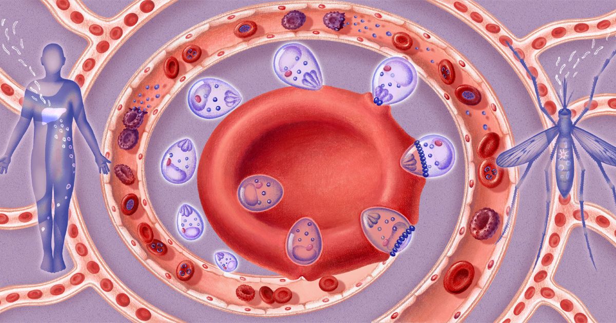
To invade red blood cells, the deadly protists create temporary molecular portals that disappear behind them — doors we can now see in fine detail. Plasmodium, the parasite that causes malaria, invades and proliferates inside red blood cells. Researchers imaged the proteins (dark purple ovals) it places on the cell membrane that let it slip through unnoticed. Ada Zejun Shen for Quanta Magazine Introduction In medieval Italy, it was accepted wisdom that anyone walking near a swamp risked falling ill from “bad air” — mala aria. Victims were struck with fever, headache, nausea, and chills, and occasionally the illness escalated to vomiting, jaundice, and death. Today, we still call this affliction malaria. For much of human history, it has ravaged populations in humid tropical and subtropical regions, killing untold numbers. In the 20th century, it was responsible for up to 5% of deaths worldwide; it still kills approximately 600,000 people each year. What malaria was and how it infected humans was deeply mysterious until barely 150 years ago. In 1880, the French physician Charles Louis Alphonse Laveran peered at a sample of a soldier’s blood through a crude microscope and discovered the true cause of the disease: not miasma, but a genus of shape-shifting single-celled protists called Plasmodium. Since then, scientists and doctors have described in precise detail the parasites’ life cycle, hosts, and disease progression, and even the nuances of various Plasmodium species, which present slightly different forms of the disease. But some crucial aspects have remained a mystery. For example, Plasmodium invades human red blood cells as part of its life cycle. To get in, the microbe creates a ring-shaped structure called the moving junction, which briefly creates a gap in the cell membrane it can slip through. The gap then disappears without a trace. It opens and closes so rapidly, at such a small scale, that imaging technologies couldn’t capture it. Researchers had no clue what the portal structure actually looked like. “Because of that,” said Chi-Min Ho, a microbiologist and structural biologist at Columbia University, “we didn’t really know anything about how it works.” Now, Ho’s lab has imaged the moving junction right at the moment of invasion, in unprecedented detail. Her team found that when the Plasmodium parasite installs molecular gaskets in the host cell, it radically remodels the cell membrane to slip through without a trace. This knowledge could help inform the next generation of malaria treatments and influence the drug-discovery process for other parasitic diseases, such as cyclosporiasis and toxoplasmosis. “It’s a totally mind-blowing paper,” said Jake Baum, a parasitologist at the University of New South Wales, who was not involved in the research. “At the end of the day, this is not molecular biology. This is biophysics.” Home Remodeling The history of malaria research has paralleled advances in microscopy, from the development of simple light microscopes like the one Laveran used to the cutting-edge cryo-electron techniques of Ho’s lab. This is because the cells the microbes invade are extremely tiny — and the parasites are even smaller. At about 8 micrometers across, red blood cells are among the smallest cells in the human body, and all species of Plasmodium are a fraction of that size during the invasion stage. Attempts to image the parasites in the act of breaking and entering got off the ground in the 1970s, when pioneering microbiologists first applied electron microscopy. These scientists observed a distinct thickening of the cellular membrane near the parasite’s access point. Then the tiny, round Plasmodium seemed to simply merge with the cell, like a water droplet coalescing into a larger pool. It would be decades before techniques developed enough to isolate the proteins that make this possible, much less image them. Another hurdle is that malaria parasites have a very complicated life cycle. They are most at home in the gut of a mosquito where they can reproduce sexually. Humans are what’s known as a reservoir host — a place for the parasite to live while it waits to be taken up by an insect. “The food supply happens to be our red blood cells,” said Kirk Deitsch, a microbiologist and immunologist at Weill Cornell Medicine in New York. “That’s why they’re invading us.” To this end, the Plasmodium parasite assumes a variety of shapes and sizes as it moves through its life stages: Sometimes it’s shaped like an egg, sometimes a spindly thread, sometimes a gangly starfish. It can move through this cycle in rapid succession or hang out in one stage (the hypnozoite phase in the human liver) for years. When it enters its tiny, round merozoite stage, the parasite is ready to invade a red blood cell. Plasmodium pulls up to the scene of the crime with all the proteins and other biological equipment it needs. The entry procedure is more delicate than simply punching a hole in the host cell. “If you’re trying to push an orange into a balloon, the balloon mustn’t pop, right?” Baum said. Based on previous research, scientists knew the broad strokes of this process. The parasite approaches a host red blood cell and inserts a ring of protein anchors into the cell’s membrane. Collectively these anchors are known as the moving junction. Plasmodium locks into them with proteins expressed on its surface — and then pushes forward. The moving junction forms a continuous portal in the host cell membrane for the parasite to travel through. Once the parasite is fully inside, the cell membrane seals up behind it. The whole process takes less than a minute. Ho and the other authors of the new paper used cryo-electron microscopy to probe the four individual protein units in each anchor. What they learned allowed them to propose a more detailed model for how the moving junction actively remodels the host cell’s membrane. The parasite injects into the red blood cell three proteins, which form a dense wedge beneath the membrane surface; a fourth protein connects this structure to the invader. Plasmodium places many of these anchors on the red blood cell surface. The proteins produce electrostatic forces and pressure in the cell membrane, which crowd and displace its lipid molecules. This deforms the membrane and reverses its curvature. It bows slightly inward around the parasite — in fact, some lipids slough off and reassemble into a protective membrane for the invader — before re-forming and sealing behind it. “It’s a really nice example of how a field has built step by step by step toward a really powerful observation,” Baum said. Once inside, Plasmodium approaches its remodeling work more comprehensively. Red blood cells are essentially bags of hemoglobin; they have no nucleus or organelles to speak of. So Plasmodium builds from the ground up. It secretes proteins that allow it to feed on hemoglobin, remove waste, and stretch the cell’s cytoplasm. Ho compared it to the conversion of an empty warehouse floor into a stylish lofted apartment. “It’s putting in plumbing,” she said. “It’s putting in the floors. It’s literally building everything for itself.” In this process, the parasite also redecorates the cell’s exterior. It is equipped with genes that allow it to make a series of distinct but closely related adhesive proteins that it can express on the red blood cell’s surface. These proteins cause the cells to stick to the walls of the blood vessels around them — one of the main causes of malaria’s painful symptoms. In 2025, research from Deitsch’s lab found that by periodically switching between proteins, the parasite can avoid tipping off the immune system to its presence for days or weeks at a time. Such extensive exterior cellular remodeling serves a purpose: It helps Plasmodium avoid a trip to the spleen, an organ that filters out dead, defective, or broken red blood cells as well as any undercover parasites. Protrusions called knobs cover the surface of an infected red blood cell. Plasmodium, the parasite that causes malaria, expresses these proteins after it invades the cell. They make cells clump to the walls of blood vessels, causing the disease’s most dangerous symptoms including organ distress, seizures, and coma. Rick Fairhurst and Jordan Zuspann, National Institute of Allergy and Infectious Diseases, National Institutes of Health The parasite itself undergoes a radical change inside the cell. It transforms from a roundish merozoite into an amoebalike trophozoite. It grows several cytoplasm “arms” and flattens out, said Christiaan van Ooij, a molecular parasitologist at Keele University in the United Kingdom. This also helps it evade the immune system. The trophozoite shape is more flexible than that of the spherical merozoite, and the immune system clocks supple cells as healthy and stiff cells as dead. The parasite then replicates asexually for about 48 hours, dividing inside the red blood cell until it’s so full of Plasmodium that it bursts. Like house flippers, the parasites move on to other blood cells. Within days, millions could be lurking in the bloodstream. In this way, malaria can quickly overwhelm a human host. It is all enabled by the moving junction that allows the parasites to set up shop without tipping off the authorities. The structure of the moving junction could help researchers intervene, with drugs to block the parasite from invading a host blood cell in the first place. Moving Target Malaria treatment has long been something of a hit-or-miss proposition. Without a granular view of how Plasmodium interacts with host cells, researchers had to test various compounds by trial and error. If a given drug seemed to fight the illness, they had little idea what part of the infection process it had disrupted. “All that work was done essentially blind,” Deitsch said. This has made developing new malaria treatments notoriously difficult. The former workhorse antimalarial drugs, such as chloroquine, sulfadoxine, and pyrimethamine, are now known to target the parasites in the bloodstream. But the Plasmodium species that infect humans are rapidly evolving resistance to these compounds. Invasion offers a tantalizing choke point for new medicines. “If you block invasion, then you stop the parasite in its tracks,” Ho said. To make her point, her team used an AI-powered protein-design platform to craft an inhibitory protein that could block the moving junction. In test tubes, it prevented Plasmodium from entering red blood cells. But its safety and efficacy haven’t been tested in animal models. It’s possible that the moving junction could be something of a skeleton key that could be used to treat other parasitic diseases. Plasmodium is a member of the apicomplexans, a large phylum of parasitic protists that also includes Toxoplasma gondii, a cat parasite, and Cyclospora, which causes explosive diarrhea. These single-celled organisms appear to enter using the same biochemical equipment as Plasmodium. “All of these are parasites that have this invasion machinery,” Deitsch said. The study of those conditions could in turn inform malaria treatment. A 2026 paper described the mechanism by which Toxoplasma senses the right moment to deploy its moving junction, and malaria may operate the same way. But the moving junction may prove to be too much of a moving target. “Some people have always argued that invasion is a really bad drug target because it’s a very short process,” van Ooij said. Any drug with the ability to stop invasion would have a roughly 60-second window in which to do so, which means it would need to remain stable in the body for a long time or act extremely quickly, or both. Ho’s team already has their sights set on another target. They plan to use similar cryo-electron microscopy techniques to solve the structure of other protein complexes involved in malaria infection, such as those adhesive proteins that keep infected blood cells from journeying to the spleen. “We’ve caught one white whale,” Ho said. “So we want to go after the other remaining white whales.” Editor’s note: This research was funded in part by the Simons Foundation, which also funds this editorially independent magazine. Simons Foundation funding decisions have no influence on our coverage. The Quanta Newsletter Get highlights of the most important news delivered to your email inbox Also in Biology Comment on this article Next article See Inside Cells Like Never Before in These Vivid Microscopic Images
Malaria Parasite Caught in the Act of Breaking and Entering
Full Article
Original Source
Read the full article at Quantamagazine →KhanList aggregates and links to publicly available news content. We do not host full articles from third-party sources. Always verify important information with original sources.