
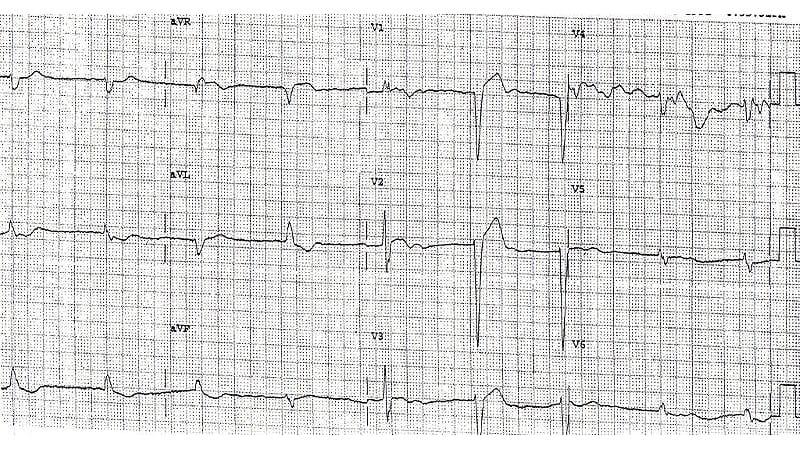
A 76-year-old woman with known atrial fibrillation, being treated with a beta-blocker for rate control, presents to her cardiologist for a routine follow-up. Her heart rate is slower than usual, and a 12-lead ECG is obtained. The patient has no symptoms.Figure 1. Courtesy of Philip J. Podrid, MDThe correct diagnosis is atrial fibrillation with right and left bundle branch block (bilateral bundle branch block) (Figure 2).Figure 2. Courtesy of Philip J. Podrid, MDDiscussionThe rhythm is irregularly irregular (although the first two RR intervals appear to be regular). Only three supraventricular rhythms are irregularly irregular. These are:Sinus arrhythmia: one P wave morphology and a stable PR intervalMultifocal atrial rhythm or multifocal atrial tachycardia: three or more different P wave morphologies and PR intervals with no single P wave morphology being dominantAtrial fibrillation: no organized atrial activityNo organized P waves are seen, but the baseline of V1 has a low-amplitude undulation; therefore the underlying rhythm is atrial fibrillation. The QRS complexes are wide (0.12 sec) and have two different morphologies. Complexes 1, 2, 3, and 5 (^) have a right bundle branch block morphology with a broad R wave in lead V1 (→) and aVR and a broad terminal S wave in lead I (←). These QRS complexes have a right axis between +90° and +180° (negative QRS complex in lead I and positive in lead aVF). Causes of a rightward axis include: Right ventricular hypertrophy (associated with a tall R wave in lead V1 and often P pulmonale)Wolff-Parkinson-White pattern (short PR interval and delta wave)Lateral-wall myocardial infarction (a deep Q wave in leads I and aVL)Right-left arm lead switch (negative P and T waves in leads I and aVL and positive QRS complex, P and T waves in lead aVR)Dextrocardia (reverse R wave progression in leads V1-V6)Left posterior fascicular block, which is a diagnosis of exclusion when no other cause for the right axis is noted.This is a case of left posterior fascicular block. The fourth, sixth, and seventh QRS complexes (+) have a left bundle branch block morphology with a deep QS wave in lead V1 (↓). The presence of both right and left bundle branch block is termed “bilateral bundle branch block” and is one manifestation of trifascicular disease.Philip Podrid, MD, is an electrophysiologist, a professor of medicine and pharmacology at Boston University School of Medicine, and a lecturer in medicine at Harvard Medical School. Although retired from clinical practice, he continues to teach clinical cardiology and especially ECGs to medical students, house staff, and cardiology fellows at many major teaching hospitals in Massachusetts. In his limited free time he enjoys photography, music, and reading.
ECG Challenge: AFib Patient With a Slow Pulse
Full Article
Original Source
Read the full article at Medscape →KhanList aggregates and links to publicly available news content. We do not host full articles from third-party sources. Always verify important information with original sources.