
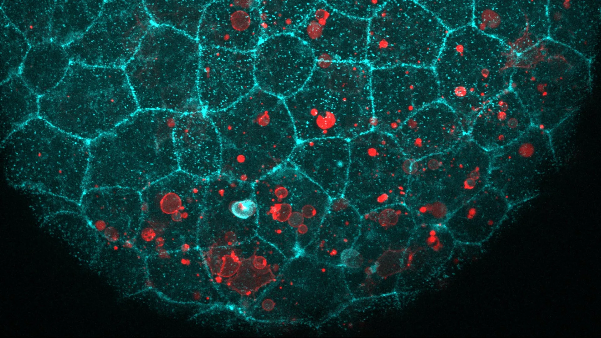
A protein best known for helping cells and tissues stay connected has another unexpected role. Researchers have found that it also helps epithelial cells, which form continuous sealed layers throughout the body, engulf nearby dead cells. The discovery could have implications for chronic inflammation. Debris from dying cells is a major contributor to inflammatory responses, so understanding how tissues remove that material may reveal new clues about what happens when the cleanup process fails. The study, published in Nature Communications, focuses on the E-cadherin complex. This molecular system includes E-cadherin along with three additional proteins. Together, they connect epithelial cells lining areas such as the skin, gut and airways, giving tissues the structural strength they need to remain intact. In these tissues, each cell connects to E-cadherin molecules on neighboring cells. Cellular Glue Takes on a Cleanup Role A team led by Verena Ruprecht examined epithelial tissues in living zebrafish and mouse embryos. They found that the same molecular machinery also gathers at the exact location where a dying cell comes into contact with the tissue. The researchers wanted to know whether E-cadherin and its partners were attaching to the dying cell in the same way they normally attach neighboring epithelial cells. To test that possibility, they carried out two experiments. First, they presented the tissue with dying cells that had been stripped of E-cadherin. The epithelial tissue removed those cells just as effectively as it removed normal dying cells. Next, the team introduced fat droplets that contained no protein at all but carried a signal normally displayed on the surface of dying cells. The epithelial cells engulfed those droplets as well. "We were intrigued to find out that epithelial cells repurpose their molecular adhesion machinery -- the 'glue' that normally holds them together -- to engulf dying cells," says ICREA Research Professor Verena Ruprecht, senior author of the study. How Cells Eat Without Breaking the Barrier Engulfing something roughly the size of another cell presents a difficult mechanical problem. Epithelial cells are tightly packed into barriers that often need to remain sealed, even while individual cells reshape themselves to remove debris. Live imaging revealed how they accomplish this. The upper and lower surfaces of the same epithelial cell can behave differently from one another. The lower surface stretches and bends around the dead cell, while the opposite side remains relatively unchanged. That upper surface, which may face the outside environment or an open space such as the inside of a lumen, continues maintaining the tissue barrier. Measurements taken before, during and after engulfment showed that the area of the upper surface changed very little. By contrast, the lower surface underwent substantial deformation during the 'eating' process. Ruprecht compares the behavior to a row of dancers standing with their arms linked. Their upper bodies remain steady while their feet perform increasingly complicated movements when a dying cell appears. "It's the same dancer with a different choreography," she says. A Molecular Rope and Brake The researchers also examined the mechanics that allow the cells to perform this cleanup. One protein in the E-cadherin complex acted much like a rope. It connected the molecular assembly to the cell's internal skeleton, allowing force to be transmitted across the surface of the material being engulfed. When cells lacked this tethering protein, or lacked the specific region that attaches it to the skeleton, they could no longer swallow dead cells. Another component behaved more like a brake on the cell's contractile machinery. Surprisingly, removing this brake did not make the cleanup process more effective. Instead, the cell became too stiff and lost its ability to properly remove dying cells. The Same Mechanism Appears in Mammals The team next investigated whether the process extends beyond zebrafish. In early mouse embryos, blocking E-cadherin caused dying cells to remain uncleared. This matched the results seen in zebrafish and suggests that the mechanism is shared among vertebrates. The new work builds on earlier research from Ruprecht showing that embryos can use epithelial tissues to cooperatively remove dying cells. That behavior represents a form of early innate immune defense. Embryos are particularly useful for studying these events because they are transparent. Researchers can observe living cells and tissues directly at a level of detail that cannot currently be achieved inside the human body. Could the Same Process Work in Adult Tissues? An important question remains unanswered. Researchers do not yet know whether this same E-cadherin dependent mechanism operates in adult zebrafish or mice, or in any type of human tissue. There are reasons to think it could. Epithelial tissues in adults are already known to remove dying cells in places including the retina, colon, airways and mammary gland. E-cadherin is also found throughout epithelial tissues in the body, and its structure has remained remarkably similar across species. Those characteristics make it a strong candidate for a more broadly used cleanup mechanism. The potential medical importance comes from what can happen when dead cells are not removed efficiently. Dying cells that remain in tissues can eventually rupture and release their contents, contributing to chronic inflammation. The findings suggest that successful cleanup depends on more than receiving the correct chemical signal to engulf apoptotic debris. Cells must also be physically capable of changing shape, applying force and wrapping around dead material without compromising themselves or the surrounding tissue. "Studying the mechanisms of how dying cells can be removed efficiently from tissues is of very high relevance to human health," concludes Ruprecht. The work was led by joint first authors Hanna-Maria Häkkinen, Marta Batet Palau and Laura F. Bianchi and supervised by Verena Ruprecht. It was funded by the Spanish Ministry of Science and Innovation, the Human Frontier Science Program, the European Union's Horizon Europe program, and the "la Caixa" Foundation, with additional support from the European Social Fund. It made use of the CRG Core Facilities for Advanced Light Microscopy, Tissue Engineering and Protein Technologies.
The “glue” holding your cells together has a surprising second job
Full Article
Original Source
Read the full article at Sciencedaily →KhanList aggregates and links to publicly available news content. We do not host full articles from third-party sources. Always verify important information with original sources.