
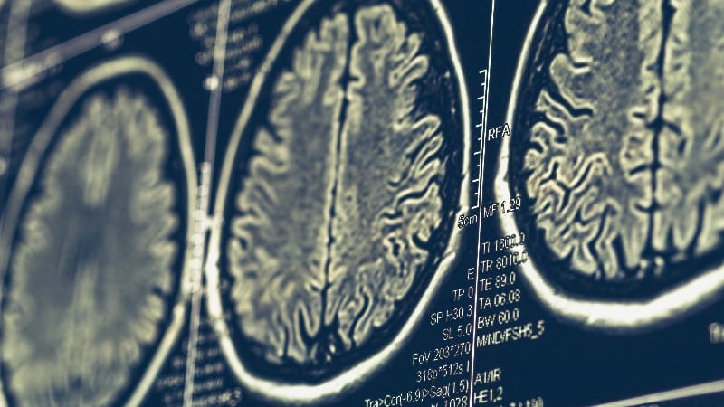
TOPLINEBaseline cortical atrophy within a specific signature region of early-onset Alzheimer's disease (EOAD) detected on MRI predicted progression from mild cognitive impairment (MCI) to dementia in patients with EOAD.METHODOLOGYResearchers conducted a longitudinal cohort study using data from the Longitudinal Early-Onset Alzheimer's Disease Study, including 130 participants aged 40-64 years (mean age at first scan, 59.59 years; 66 men) with EOAD at the MCI stage (Clinical Dementia Rating [CDR] global score, 0.5) and 97 cognitively normal, age-matched control individuals (mean age at first scan, 56.85 years; 35 men).Participants underwent structural MRI and amyloid PET imaging at baseline.The primary outcome was time from baseline to progression to dementia, defined as the first follow-up visit at which a participant met the criteria for a CDR global score of 1.Researchers assessed the association between baseline cortical atrophy within the EOAD-signature region (derived from a previous study identifying cortical areas that showed greater atrophy in patients with EOAD compared with cognitively normal participants) and the risk for progression to dementia.The mean follow-up duration was 21.73 months. The base model was adjusted for age, sex, and baseline CDR Sum of Boxes score, while the extended model additionally adjusted for the EOAD-signature W-score.TAKEAWAYAbout 65% of patients with EOAD progressed from MCI to dementia. The magnitude of baseline cortical atrophy within the EOAD-signature region predicted the time for progression (hazard ratio [HR], 1.24; P < .0026). An increase in the magnitude of atrophy by one SD was associated with a 24% higher risk for progression.Baseline cortical thickness across the whole cortex (HR, 1.31; P < .0026) and within each brain network except the somatomotor network (HRs ranging from 1.22 to 1.33; all P ≤ .0101) predicted progression to dementia, but average hippocampal volume did not.Atrophy in the EOAD-signature region yielded the highest sensitivity (lowest Akaike Information Criterion value) in predicting progression, followed by atrophy in the frontoparietal control network.The extended model incorporating EOAD-signature W-score showed a better predictive performance than the baseline model (P = .011).IN PRACTICE"Leveraging a relatively simple and cost-effective MRI-based measure, which is typically possible to obtain from scans collected in routine MRI protocols during diagnostic assessment, could facilitate the creation of individualized prognostic and monitoring strategies in clinical practice and in clinical trials," the authors wrote.SOURCEThe study was led by Thiago Paranhos, Massachusetts General Hospital and Harvard Medical School, Boston. It was published online on August 26 in Neurology.LIMITATIONSThe model was developed and validated within a single cohort that was predominantly non-Hispanic White and included mostly patients with amnestic MCI, limiting the generalizability of the findings.DISCLOSURESThe study was funded by the National Institute on Aging and the Alzheimer's Association. The authors reported no relevant conflicts of interest.This article was created using several editorial tools, including AI, as part of the process. Human editors reviewed this content before publication.
Cortical Atrophy Signals Progression in Early-Onset AD
Full Article
Original Source
Read the full article at Medscape →KhanList aggregates and links to publicly available news content. We do not host full articles from third-party sources. Always verify important information with original sources.